
Deep in the bowels of the National Gallery of Art’s West Building, one painting—an icon, about three square feet in size, of a calm-eyed saint—rests in an unusual frame. Instead of hanging on a clean gallery wall, it is fixed in the center of a complex, room-sized scaffold. A few feet away are a bank of computers and a complicated apparatus about the size of a coffee table: a neat assemblage of metal boxes, cables, lights and lenses, looking like the stacks of equipment in an avid TV-watcher’s entertainment center.
The scene looks more like something from a NASA scientist’s laboratory than a museum conservation and restoration workshop—and that’s understandable. This room is the research headquarters of a team that includes Professor Murray Loew of the George Washington University School of Engineering and Applied Science (SEAS). Here, with the help of a five-year, $500,000 grant from the National Science Foundation, scientists have been working to develop imaging tools that can peel back the centuries, using an infrared spectrometer to map an artist’s materials, techniques and even artistic process.
“Our goal,” Dr. Loew said, “was to build a camera that can see beneath the surface, so to speak, of a painting.”
Painting analysis historically has been done point-by-point, using spectrometry—studying the way substances reflect light at different narrow bands of frequency—to figure out what materials make up small samples of a work. That’s necessary knowledge for conservators, who need to understand the artist’s materials in order to care for and restore paintings.
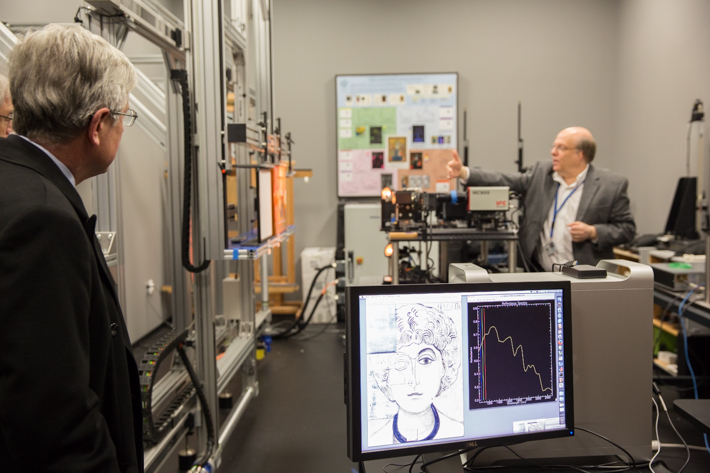
With this new instrument, however, conservators can see what they are working with on a larger scale. The spectrometer scans the painting as a whole, creating high-resolution “media maps” of the different binders, pigment materials, sealants and varnishes that create it. Then, with the help of sophisticated mathematical models, these maps can be overlaid on one another to create a complete, three-dimensional picture of a painting’s many layers.
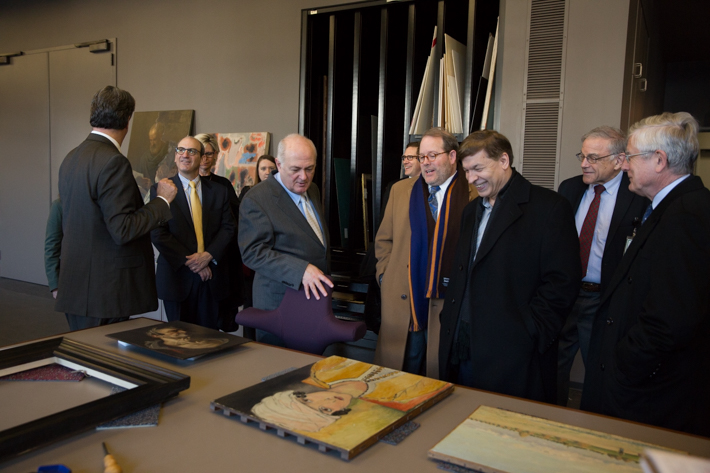
Dr. Loew invited George Washington President Steven Knapp and SEAS Dean David Dolling to take a behind-the-scenes look at the team’s research-in-practice at the National Gallery of Art last Wednesday. George Washington Today went along for the tour.
The project spans multiple disciplines, bringing together art historians, computer scientists, chemists and conservators, among others.
“We use advanced analytical macro-scale imaging methods—techniques developed by NASA and the like to explore and map minerals on planets—and [apply them] to paintings,” explained John Delaney, senior imaging scientist in the NGA’s scientific research department and a co-principal investigator with Dr. Loew on the NSF grant.
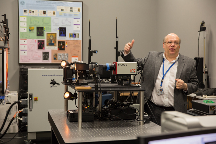
John Delaney demonstrates the camera-scanner with which he and Dr. Loew conduct their research. (Photos: William Atkins)
The camera-scanner that he and his team have used in their research—that table-sized arrangement of electronics that dominates the center of the laboratory—was based on sensor technology purchased from a division of Lockheed Martin. But it had to be adapted to the demands of art.
“Paintings want to be at a much lower light level,” Dr. Delaney explained. “So we had to improve the sensitivity of these cameras by at least an order of magnitude.” The new sensor, cryogenically cooled to reduce noise, provided that increased sensitivity.
The process can reveal more than just chemical makeup. By mapping a substance like graphite, for instance, analysts can see the sketch beneath the painting, revealing the changes that an artist or his workshop made between draft and final work.
Barbara H. Berrie, head of the scientific research department at the National Gallery of Art, displayed an analysis of 14th century Flemish painter Jan Van Eyck’s “Annunciation” to demonstrate. Picked out by the spectrometer was a detailed graphite underdrawing of the sort for which Van Eyck was famed. The painter appeared to have corrected some anatomical choices between sketch and final work: Mary’s ear, for instance, had moved upward on her skull.
Even more compelling, Dr. Berrie said, was the iconography of a stained glass window, high in the background, which appeared to have changed. Those symbolic alterations could give insight into the painter’s decision-making process.
“This [media map] becomes a primary document for the artist’s workshop methods,” she said.
“Some of the techniques we have developed are based on work we’d done before in medical imaging,” said Dr. Loew, whose background is in biomedical engineering. “The idea of registering—of overlaying images accurately—is important in medicine too, where you have different kinds of images communicating different kinds of information: x-rays, MRI and CT and so on. In surgical planning, for example, it’s important to line those up very accurately.”
The same principle, he said, can be applied to art. “It’s a little bit like planning for a non-invasive procedure on the painting,” Dr. Loew said.