
A team of researchers at the George Washington University has developed a faster method to predict whether potential new drugs will cause heart arrhythmias using optogenetics, a technique that uses light to control cells.
The research team used light to both make the cardiac cells beat and to optically measure their response, allowing the scientists to automate the drug-testing process and to rule out potentially dangerous drugs faster.
The researcher’s new system can test 30,000 light-responsive cells in less than 10 minutes—a far shorter timeline than the existing standard practice, which can take years.
The technique streamlines what has been a primarily manual process that researchers carry out to comply with Food and Drug Administration testing requirements and ensure the safety of drugs.
“This new method has the potential to vastly improve the speed at which we get safe and essential drugs to seriously ill patients,” said Emilia Entcheva, professor of biomedical engineering at GW and senior author of the paper.
“Importantly, we demonstrate this can be done using a patient’s own blood cells by combining stem-cell techniques with our new method. One of the biggest challenges with developing new medications is testing to make sure we are treating a problem without causing any cardiac toxicity. This technology helps us to ensure that we are doing just that.”
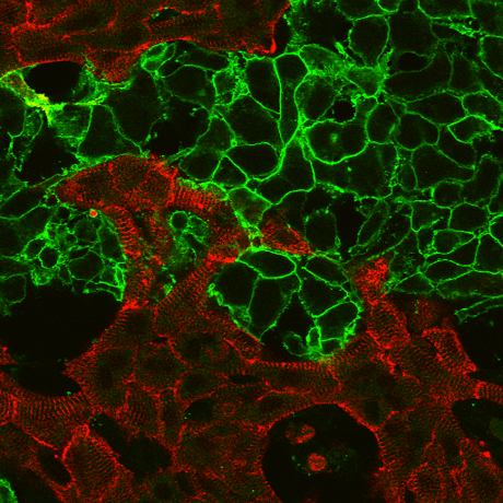
Optogenetics has been used in neuroscience for a decade, but this technique is relatively new in cardiac research.
The research is outlined in a paper, “OptoDyCE as an Automated System for High-Throughput All-Optical Dynamic Cardiac Electrophysiology,” which published in Nature Communications on Tuesday.
In order for medications to be approved by the FDA, they must be vetted to make sure they do not pose a danger to the heart or any other part of the body.
In the earliest phases of testing, cells are treated with the medication to see how they react. Traditionally, the most reliable method is to measure their response manually, by directly sticking probes into the cell. New high-throughput technologies speed up this practice. However, until now no such high-throughput system has been available for work with heart cells.
Dr. Entcheva’s research helps provide a more complete picture of how the drug being tested could affect the heart early in the development process, saving time and resources that can be spent on other new drug candidates.
Aleks Klimas, first author on the paper and Dr. Entcheva’s Ph.D. student, said the new system not only allows for faster testing but also provides a safer way to do measurements when using hazardous materials.
“The benefit of optical stimulation and optical recording is that it provides a way to dynamically control millions of cells simultaneously without needing to come into contact with the sample,” Ms. Klimas said.
Separate from its uses in the drug discovery arena, the method also can be used by scientists to confirm that stem cells have properly matured into heart cells. The new technique will help improve the quality of these newly created heart cells and speed up their deployment in the clinic.
The technology has the potential to be adopted by pharmaceutical companies to expedite the drug development process. Following the completion of her Ph.D. in biomedical engineering at GW, Ms. Klimas hopes to commercialize the idea and develop a business to market the device.
Other authors on the paper are: Christina M. Ambrosi at GW and former members of Dr. Entcheva’s lab at Stony Brook University: Harold Bien, John C. Williams and Jinzhu Yu.